What can 12 minutes of fMRI tell you about your fetus’s brain?
Author: Lavinia Carmen Uscătescu
Editors: Elisa Guma, Elizabeth DuPre, Kevin Sitek
Lay summary of publication by Ji et al (2022): “Fetal behavior during MRI changes with age and relates to network dynamics”
That fetuses move inside the womb is nothing new, but how these early movement patterns could predict neonatal health is only beginning to be understood. So far, generally reduced intra-uterine movement has been associated with preterm birth and mild language delay, while more active fetuses have shown enhanced neonatal brain development. It could therefore prove useful to learn whether specific fetal movement patterns could be used as early indicators of potential developmental delays.
Researchers at the New York University School of Medicine have set out to decode the hidden information in fetal movement patterns. Their paper, led by Dr. Lanxin Ji, “Fetal behavior during MRI changes with age and relates to network dynamics”, was awarded the Human Brain Mapping Editor's Choice Award at the 2023 OHBM Annual Meeting. Check out our interview with Dr. Ji here!
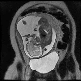
For this study, the researchers recruited healthy individuals that were six months pregnant and obtained functional MRI scans of their fetuses. This 6 month time point was chosen because there is evidence to suggest that a fetus’s brain begins to show signs of intrinsic connectivity (i.e., when brain areas spontaneously communicate to each other) at this stage in development. The researchers were especially interested in the dynamic fluctuations of co-activation patterns in a brain region called the supplementary motor area, thus aiming to isolate brain networks that support motor behavior of fetuses. This area is part of the frontal cortex and is functionally involved in the motor control network, mainly in motor planning and learning. To quantify fetal motion, the researchers employed a deep learning-based automated tool to extract fetal brain from maternal tissues and used a measure called framewise displacement—a motion parameter which is quantified at each time point throughout the fMRI scan.
After birth, the researchers assessed the motor development of the infants at 7 months and then at 36 months of age. They showed that persistence of a specific co-activation pattern of these motor networks in the 6 month fetus correlated with newborn motor development at 7 months of age.
The relevance of these findings is twofold. First, motion during fMRI scanning is usually discarded as a mere artefact that just adds noise to the signal, so finding that it may in fact carry useful information is remarkable. Second, it is noteworthy that early fetal behavior and brain activity can in fact anticipate later developmental milestones. This raises hope that perhaps, with enough refinement and standardization of this approach, we may not be far from identifying very early signs of delayed development and therefore prompt timely intervention.
Original Research: Ji, L., Majbri, A., Hendrix, C.L., Thomason, M.E., (2023). Fetal behavior during MRI changes with age and relates to network dynamics. Human Brain Mapping, 44(4): 1683-1694. doi: 10.1002/hbm.26167
