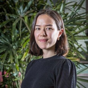
Interview with Justine Y Hansen, winner of the Karl-Zilles Award in 2023
Author: Simon Steinkamp
Editors: Elisa Guma, Elizabeth DuPre, & Kevin Sitek
Interview with Justine Y Hansen, winner of the 2023 Karl-Zilles Award
Next up in our award-winner interview series is Justine Y. Hansen, who received the 2023 Karl Zilles Award in Integrative Neuroscience in 2023. This award was introduced at the OHBM 2022 annual meeting in Glasgow in memory of Karl Zilles, to honor his pioneering work integrating modern neuroanatomical approaches with multi-modal in-vivo neuroimaging. The award series recognizes PhD students and postdoctoral fellows who are continuing in this tradition, developing new and innovative approaches for examining neuroanatomy
Justine Y. Hansen does just that. In her impressive work, she thoroughly investigates how neurotransmitter distributions shape cortical architecture across many modalities and spatial scales. She led a huge collaborative open science effort to collate positron emission tomography (PET) data across research groups, resulting in data from more than 1200 healthy individuals, mapping 19 different receptors and transporters across 9 neurotransmitter systems. Using multiple imaging modalities, such as functional and diffusion magnetic resonance imaging (fMRI, dMRI) and magnetoencephalography (MEG), she shows that the neurotransmitter receptor density maps follow known structural and functional organizational principles. Further, she shows that the receptor density maps are associated with patterns of cortical atrophy across 12 different psychiatric and neurodevelopmental disorders.
This study, published in Nature Neuroscience, has been widely recognized and was awarded a Brain Star award by the Canadian Association for Neuroscience in 2022. Justine extended this work to examine how cortical maps of gene expressions, neurotransmitter identity, metabolism, and myelination relate to cortical abnormalities in 13 neurodevelopmental, neurological, or psychiatric conditions (published here; check out the lay summary of this article on our blog!). She further leveraged gene expression and receptor density mapping (using PET or autoradiography) to assess whether gene expression measures can be used to estimate neurotransmitter receptor/transporter densities in the cortex (in this publication). Finally, in her newest work, she investigated how the brainstem, an often overlooked part of the brain, integrates into the larger cortical architecture.
Justine completed her bachelor’s degree in Neuroscience in 2020 at McGill University and is currently a PhD student in the Network Neuroscience Lab led by Bratislav Misic at the Montreal Neurological Institute. We are grateful that Justine was willing to answer a few questions about her work and research trajectory. Read on to learn more!
1. How did your passion for neuroscience start?
Justine Y Hansen (JYH): Doesn't everyone think the brain is cool?
2. What have been the most important factors that shaped your scientific trajectory up to this point?
JYH: I am hugely grateful for and indebted to amazing scientists and mentors along the way who have served as role models and helped (and currently help) me shape what type of scientist I want to be. That's a bit vague, but honestly my scientific trajectory is still in its infancy so we'll see what happens in the future… .
3. How can researchers and clinicians make use of the impressive contributions you have made to the field of brain mapping?
JYH: One main theme in my work is that combining brain phenotypes across different spatial scales (and therefore different datasets, data modalities, and people) can help us understand how the brain is integrating all these layers of information. Both the brain maps as well as the methods that I use and help share will hopefully facilitate future multimodal, multiscale analyses of the human brain. I think the PET receptor images in particular will be useful to clinicians and researchers alike because these neurotransmitter receptor systems are fundamental to both the link between structure and function, as well as many diseases and disorders.
4. What were the biggest challenges you faced in integrating modalities across space and scale (i.e., neuroimaging, PET, gene expression, etc.)?
JYH: One big challenge is that it's impossible for me to be an expert on every data modality, so there is always plenty I don't know about the data I'm working with. Although this can feel overwhelming at times, it's also a great opportunity to collaborate with the experts. I feel extremely grateful and lucky to have learned so much from individuals who are truly experts in their field (whether that be image preprocessing, or biological mechanisms of neurotransmitters and receptors, or open science practices, etc).
Another challenge comes down to the methodology. How do we compare measurements that are capturing processes on completely different spatial and temporal scales, and from different people? Another consideration is that they are all group-averages, so they may not even be a good representation of the underlying biological mechanism (?!). I try to conduct extensive sensitivity and robustness analyses but this is an ongoing challenge and a big direction for future work; that is, better methods and data for integrative analyses across data modalities and scales.
5. What is the next big innovation we need in the field of multi-modal in-vivo imaging for the study of neuroanatomy and brain function?
JYH: As much as I am a big fan of big data, I think the next big thing is going to be densely sampled individuals. How do all these markers—anatomy, structure, function, molecular profiles, gene expression, metabolism, you name it—relate to one another in a single individual? I would love to know :)
